
Market promotion of medical devices
In today's rapidly developing medical industry, patient safety has always been the core destination of all work, and safeguarding safety requires two key supports - solid clinical competence of medical staff and continuously optimized medical quality and safety system. The high simulation training driven by 3D printing technology is becoming an important link connecting these two supports, bringing revolutionary changes to medical education and patient safety protection.
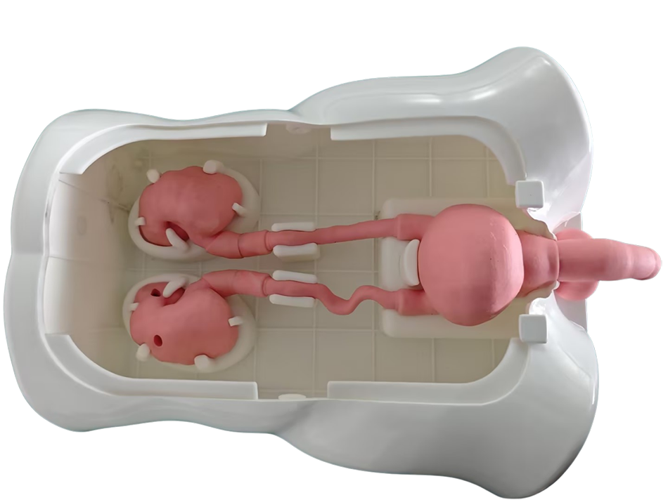
Recently, a multi material 3D printed urinary endoscope training model has been released. It is more in line with the anatomical characteristics of the urinary system, upgrading its functions and bringing innovation to urology training, becoming a practical tool for clinical teaching.
01 Technical Core:
Medical simulation is no longer a simple "training tool", but a core methodology that runs through the cultivation of medical talents and the improvement of medical quality. Through the dual track approach of CBE (Competency Education) and HQI (Healthcare Quality Improvement), 3D printed high fidelity models driven by urology, cardiology, and gastroenterology are pushing the authenticity and practicality of simulation training to new heights, becoming a typical example of various specialties practicing CBE and HQI concepts.
In the field of urology, a urinary endoscope training model based on multi material 3D printing technology has attracted much attention. The core advantage of this model lies in its precise replication and interactive experience of the human urinary system. The structural design is based on the three-dimensional reconstruction of high-precision CT and MRI imaging data, and the complete structure printing from the urethra, bladder to the upper ureter is completed in one go through multi material 3D printing technology. Unlike traditional static models, it breaks through by reproducing the true feedback of clinical operations: the resistance of the instrument in the cavity, the friction sensation through narrow areas, and the soft touch of the tissue, all of which are infinitely close to the real human body. This high degree of simulation allows the training of urologists to no longer be limited by the scarcity of clinical cases.
In the field of the heart, 3D printing technology has achieved a leap from anatomical simulation to functional replication. The 3D printed heart model is also based on high-precision medical image reconstruction, using elastic biomimetic materials to print structures such as myocardium, valves, and blood vessels. It can not only accurately reproduce anatomical details such as chamber morphology and valve opening and closing angles of the heart, but also simulate pathological states such as myocardial hypertrophy and valve stenosis. For cardiac intervention surgery training, doctors can repeatedly practice key operations such as catheter puncture and stent implantation on the model, feel the elastic feedback of the vascular wall and the resistance changes of instrument advancement, effectively reducing the risks in clinical surgery.
The 3D printing simulation models in the field of gastroenterology focus on the complex structure and dynamic functions of the digestive tract. The model covers the complete digestive tract structure such as the esophagus, stomach, and intestine, and replicates the smooth muscle movement of the esophagus, the softness of the stomach wall, and the bending characteristics of the intestine through gradient hardness materials. For the training of gastroscopy and colonoscopy operations, the model can simulate pathological scenarios such as stenosis, obstruction, and polyp attachment in different parts. Doctors can practice endoscopic navigation, polyp removal, and other skills during training to solve the pain points that traditional models cannot reproduce the dynamic physiological characteristics of the digestive tract.
02 R&D Challenge:
The seemingly perfect training model embodies the R&D team's continuous efforts to overcome technical challenges. Whether it is the soft connected structure of the urinary system, the complex chamber valves of the heart, or the curved and wrinkled shape of the gastrointestinal tract, the difficulty of 3D reconstruction far exceeds that of hard tissue printing in orthopedics and other fields. The R&D team needs to balance the adaptability of specialized instrument operation while accurately preserving human anatomical details - ensuring that the model size and shape are consistent with the human body, while also making the operation feel real and controllable.
Through precise mixing and spraying using 3D printing technology, natural transitions between different tissues have been achieved, avoiding the problem of "texture discontinuity" in traditional models. In addition, the R&D team has achieved precise matching between dynamic functions and operational scenarios through multiple rounds of optimization, while ensuring the portability and stability of external devices, ultimately forming a practical and easy-to-use solution.
03 Clinical value:
The clinical application value of this 3D printed urinary endoscope training model is mainly reflected in providing doctors with a safe, controllable, and reproducible practical training platform. The precision requirements for endoscopic surgery in urology are extremely high, such as ureteroscopy lithotripsy, pyelolithotomy, etc. Once the operation is incorrect, it may lead to serious complications such as urethral injury and massive bleeding. However, opportunities for practice on patients are limited, and traditional models cannot provide real operational feedback, resulting in slow skill improvement for doctors. With this model, doctors can confidently undergo repeated training.
The 3D printed models in the field of heart provide crucial support for training in complex heart surgeries. Surgical procedures such as congenital heart disease repair and heart valve replacement have extremely high risks and require strict precision and on-site judgment from doctors. By 3D printing a heart model, doctors can simulate the surgical path before surgery, refer to the model planning scheme during surgery, and repeatedly hone their operating techniques on the model, familiarize themselves with the spatial relationships of the heart anatomy, and significantly reduce the rate of surgical errors.
Simulation training in the gastrointestinal field is equally significant. Early gastric cancer screening under gastroscopy, colonoscopy polypectomy and other procedures require doctors to have precise endoscopic observation and operational control abilities. 3D printed gastrointestinal models can simulate different types of lesion morphology and dynamic environment of the digestive tract, allowing doctors to accumulate operational experience without patient risk, and improve lesion recognition rate and surgical success rate.
04 Market prospects:
With the increasing demand for high fidelity simulation training in medical education, urinary endoscopy training models are facing broad market prospects. According to a research report by Mordor Intelligence, the market size of 3D printed medical equipment is estimated to be $2.76 billion in 2025 and is expected to reach $6.19 billion by 2030, with a compound annual growth rate of 17.5% during the forecast period (2025-2030). High fidelity training models are one of the fastest-growing sub sectors.
In addition to urology, this technology also has great potential for extension to other specialties. The R&D team stated that in the future, endoscopic training models for more parts such as the gastrointestinal tract and respiratory tract can be developed based on the same technological framework, forming a complete series of specialized training models. In addition, the model can also be applied in the field of medical device testing, providing a realistic simulation of the human anatomy environment for the development of new endoscopic devices; In terms of patient education, it can also help patients intuitively understand the condition and surgical plan, and improve communication efficiency.
At the policy level, various provinces in China have successively issued fee items for 3D printed medical models, providing policy support for the development of the industry. Domestic medical 3D printing companies are also actively exploring diversified business models, promoting technology implementation through direct supply to hospitals, cooperation with medical device suppliers, and co building in hospital application centers. Of course, the development of the industry still needs to address issues such as establishing quality control standards and protecting patient privacy. However, with the maturity of technology and the improvement of standards, the market size of 3D printed medical training models will continue to expand.
